Contents
Share
Researchers combined electrical impedance tomography with an ion transport model to study the activity of ion channels on cell membranes
Ion transport between cells and their surroundings occurs through ion channels on the cell membrane. This process can also be anisotropic, wherein ions are distributed non-uniformly around the cells. Recently, researchers have utilized electrical impedance tomography (EIT), a non-invasive technique, along with an ion transport model to detect ion channel activity. Assessment of such transport can facilitate tissue detection and drug discovery.
Title: Electrical impedance tomography (EIT) imaging of outside-the-cell ion concentration changes associated with ion channel activation
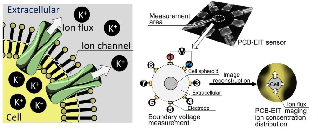
Caption: Researchers first generated a non-uniform ion distribution around a cell-spheroid. Then, they performed EIT using a printed circuit board sensor. It produced images depicting the ion distribution. An ion transport model was also utilized to calculate the associated transport coefficients.
Credit: Daisuke Kawashima of the Institute for Advanced Academic Research at Chiba University
License Type: Original Content
Usage Restrictions: Not to be reproduced without permission
The cell membrane has numerous channels for the transport of various substances, including ions, between the cell and its environment. Ion transport determines the ion exchange rate (or the transmembrane transport coefficient), which, in turn, controls biological functions, such as nerve excitation, heartbeat, muscle contraction, and hormone secretion. It can also be anisotropic, wherein a non-uniform distribution of ions causes different ion exchange rates in different directions. This effect is quite pronounced in heterogeneous tissues. Therefore, the boundaries and overlap of tissues can be detected by measuring the associated anisotropic transmembrane transport.
Recently, a group of researchers, led by Daisuke Kawashima, an Assistant Professor at the Institute for Advanced Academic Research at Chiba University, has measured anisotropic transmembrane transport by modifying the EIT technique and improving the ion transport model. Their work was published in Volume 34, Number 3 of the Measurement Science and Technology journal on 22 December 2022. It is co-authored by two Chiba University professors—Masahiro Takei of the Laboratory on Multiphase Flow and Visualization at the Graduate School of Engineering and Takeshi Murata of the Laboratory of Biostructural Chemistry at the Graduate School of Sciences.
Prof. Kawashima briefly explains the research methodology. “First, a non-uniform ion distribution was generated around a spheroid—cell aggregate that mimics tissues—by injecting two different sucrose solutions from both sides. Following that, EIT was performed using a microelectrode array sensor mounted on a printed circuit board.”
The solutions were of three concentrations, relative to the cell aggregate: isotonic, hypotonic, and hypertonic. The technique successfully produced images depicting the non-uniform ion distribution due to the anisotropic transmembrane transport. Subsequently, the researchers applied the ion transport model to calculate the associated transport coefficient and anisotropic factor. The latter was 0.34 ± 0.24 in iso-hyper, 0.58 ± 0.15 in iso-hypo, and 0.23 ± 0.06 in hyper-hypo solution combinations. The researchers verified these results by observing the fluorescence ratios of potassium ions—the most abundant species involved in cell ion transport—around the cell-spheroid. They were consistent with the EIT values for all three combinations.
“Consequently, the proposed EIT-based imaging technology provides a simple and non-invasive anisotropic transmembrane transport measurement method for cells and tissues. It can immediately measure the drug response associated with ion channels, leading to more efficient and shorter preclinical testing,” concludes Prof. Kawashima.
The model is expected to serve as a new evaluation platform for medical discovery by contributing to the realization of a rapid drug development process.
About Prof. Daisuke Kawashima
Daisuke Kawashima received the B.S., M.S., and Ph.D. degrees in Mechanical Engineering from Tokyo Metropolitan University in Japan in 2012, 2014, and 2017, respectively. In 2017, he joined Chiba University in Japan as a Postdoctoral Fellow. He is currently serving there as an Assistant Professor at the Institute for Advanced Academic Research. Prof. Kawashima has published over 40 research papers, which have been cited 250 times. His research interests include mass transfer in cells/tissues, electrical spectral analysis and electrical tomographic imaging applied to biomedical and industrial fields.
Funding Information
This work was supported by JST SPRING, Chiba University, and JSPS KAKENHI.
Reference
Title of original paper: Assessment of anisotropic transmembrane transport coefficient vector of cell-spheroid under inhomogeneous ion concentration distribution fields by electrical impedance tomography
Authors: Songshi Li1, Daisuke Kawashima1,2, Kennedy Omondi Okeyo3, Takeshi Murata4, and Masahiro Takei1
-
Affiliations:
- Graduate School of Engineering, Chiba University
- Institute for Advanced Academic Research, Chiba University
- Department of Biomechanics, Institute for Frontier Life and Medical Sciences, Kyoto University
- Graduate School of Science, Chiba University
Journal: Measurement Science and Technology
DOI: 10.1088/1361-6501/acaa4a
Contact:
Daisuke KAWASHIMA
Institute for Advanced Academic Research, Chiba University
Address: 1-33 Yayoi, Inage, Chiba 263-8522, Japan
Email: dkawa@chiba-u.jp
Public Relations Office, Chiba University
Address: 1-33 Yayoi, Inage, Chiba 263-8522, JAPAN
Email: koho-press@chiba-u.jp
Recommend
-

Examining Social Injustice of Modern Society through “Child Poverty”: Creating a Society Where Every Parent is Respected, and Every Child Thrives
2023.05.26
-
Viewing a Diverse World through the Lens of Agriculture and Food: Enriching Global SDGs Education with Insights from Extensive Overseas Field Research
2023.08.31
-
Live on the Moon, Settle on Mars: Research Center for Space Agriculture and Horticulture for Space Food Production and Supply
2023.10.20